Professor David N Furness
- Title
- Professor Emeritus of Cellular Neuroscience
- Location
- Electron Microscope Unit : 33484
- Contact me
- Try my office or arrange an appointment by e-mail.
- Phone
- +44 (0)1782 733496
- d.n.furness@keele.ac.uk
Biography
I am an award winning electron microscopist with over thirty years of experience in the principles and practical application of EM in both commercial and research sectors. My interest in microscopy began early with a birthday present of a small light microscope at 12.
Recognising its limitations, I saved up and acquired a more elaborate microscope. These microscopes established an interested in Protozoa, in particular, and so began my career in microscopy. This developed into the desire to be a researcher who used electron microscopes to study cells generally. I completed an undergraduate zoology degree at Manchester University which included Cell Biology and Protozoology courses, and did a third year project on the ultrastructure of rumen Protozoa. I obtained a First Class Honours and won the Zoology prize which was followed by a departmental PhD position to continue my study of rumen ciliates, presenting my work first at a meeting of the Royal Microscopical Society, at which I won a prize for the best young microscopist, and subsequently giving talks at UK protozoology conferences. Also during my PhD I made an early foray into immunofluorescence techniques, then in their infancy, X-ray microanalysis, and high resolution scanning electron microscopy (SEM) on a Phillips 400T TEM which was capable of in-lens imaging. This was the first high resolution imaging of protozoa certainly at Manchester and possibly elsewhere. I wrote three papers on my PhD research before submitting the final thesis and attending an international Protozoology conference in Nairobi.
Immediately after completing my PhD, I took up a post-doctoral position at Keele University, building on my EM and immunofluorescence experience, to study cytoskeletal structures after damaging the ear with strong antibiotics. This introduced me to hearing research, which I have been involved with ever since. At the end of my post-doctoral period I applied for and was awarded a Wellcome Trust lectureship (five years, with a two year extension) and with my start-up funds purchase an X-ray microanalysis system to add to Keele’s growing electron microscope unit. Our research into antibiotic damage and cytoskeletal organisation in the hearing organ continued, with a PhD student whom I co-supervised and we developed immunofluorescence microscopy at Keele and 3D reconstruction techniques to evaluate microtubular organisation in sensory cells of the inner ear. In 1994 I was given a permanent position at Keele University. I have been director of the Electron Microscope Unit (EMU) at Keele since 1995.
About that time, I led on the development of a new Neuroscience Principal Course at Keele, part of the dual honours system, and this has flourished, now boasting an intake of around 60 students p.a. I was course director for Neuroscience until my promotion to Reader in 2001, when I passed on the mantle, but I still organise several modules and teach on many more, having been nominated for student led teaching awards several times.
I was also at that time Director of the MacKay institute for Communication and Neuroscience, leading on research into hearing with grants from the MRC, Wellcome Trust and a number of hearing charities. After changes at Keele in 2006, I became theme lead for Cell and Tissue Engineering, subsequently splitting into a new theme which I led, Neuroscience and Human Metabolism, in the new Institute for Science and Technology in Medicine. Most recently I was promoted to a personal Chair in Cellular Neuroscience and became Research Director for the School of Life Sciences in 2015.
I am passing that mantle on as well in September 2020. I remain a leading researcher in hearing and hearing loss therapies, having held grants from the MRC, Wellcome Trust, BBSRC and a number of hearing charities. I am also responsible for Keele University’s Research Excellence Framework submission for Biological Sciences.
Research and scholarship
 My main research interests lie in the field of auditory neuroscience. I am presently investigating the role of fibrocytes in the cochlea. These are cells that are associated with homeostatic mechanisms that regulate the composition of cochlear fluids. These cells appear to go wrong in certain forms of deafness, including age-related deafness (presbyacusis). My group has funding from Deafness Research UK to investigate whether a stem cell/replacement cell strategy might be employed to prevent or ameliorate age-related deafness by replacing defective fibrocytes.
The main techniques we employ are electron microscope, post-embedding immunogold electron microscopy, confocal microscopy and cell culturing. We have received funding for our work from the Wellcome Trust, MRC, Deafness Research UK, Midlands Institute of Otology, Royal Society, Physiological Society, British Tinnitus Association and others.
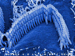
Another of my major research interests is cochlear hair cell structure, composition and function. I am focussing on the hair bundle and how the hairs (stereocilia - see picture) function to convert mechanical action from sound stimulation into electrical signals and nerve impulses in nerve fibres going to the brain.
I am also director of the Keele University Electron Microscope Unit which offers a range of microscopy facilities.
For some examples of our electron microscopy please visit the galleries on the EM Unit web pages.
Publications
Head of School
Professor Pip Beard
Email: p.m.beard@keele.ac.uk
School address
School of Life Sciences
Huxley Building
Keele University
Staffordshire
ST5 5BG
Tel: +44 (0) 1782 734414
Email: lifesciences.office@keele.ac.uk
Enquiries
Tel: +44 (0) 1782 734414
Email: lifesciences.office@keele.ac.uk
You can find information about Programme Directors on our Contact us page.
A full list of School staff is available with details of individual rooms and telephone numbers, email addresses, details of research interests and other information.