Electron Microscope Unit
We have a range of microscopic techniques we can use to capture images and acquire data from biological, geological, physical and chemical specimens. Whether it is cells and organelles, pathological samples, microfossils and rock samples, toothpaste or tiny pond creatures, we can open up a new world of investigation for you.
Overview
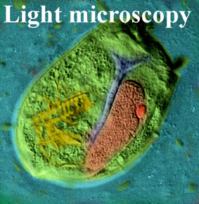 This section is intended for people who have little knowledge of microscopes and for those looking for teaching and educational resources. Teachers, if you would like more information or to book a visit to our Electron Microscope Unit, please go to the Teacher’s page
The first microscopes were made by a Dutch scientist, Anton von Leeuenhoek and Robert Hook in England in the 17th century. Like the first telescopes, they revealed a whole new world to scientists and naturalists.
From the observation of tiny pond creatures (animalcules) and the cellular structure of both plant and animal tissues, the first microscopes made startling new discoveries that have shaped our understanding not only of the microworld but also of our larger day-to-day world.
There are two basic kinds of microscope, light microscopes and electron microscopes. Did you know that whilst the smallest thing you can see by eye is about 1 tenth of a millimetre across, a typical light microscope can enable you to see things 500 times smaller and a modern electron microscope 500000 times smaller!
|
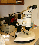
Light microscopes use glass lenses to focus light that is reflected from or passes through a specimen. They have an objective lens which forms a magnified image of the object and an eyepiece lens designed to focus the light into your eyes and magnify the image further. A zoom microscope enables you to see an object with light reflected from it at relatively low power. |
|
|
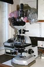
A compound microscope is designed to look at thin samples (sections) through specimens and has a source of light underneath, focussed onto the specimen through a set of lenses called a condenser. With suitable lenses, they can magnify up to several hundred times. |
|
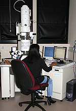
Electron microscopes (EM), surprisingly, are pretty much the same as light microscopes! They use a beam of electrons rather than light, but have objective lenses like a light microscope. The big difference is that very few things are transparent to an electron beam, unlike light. Even normal air will stop the beam very quickly and it cannot travel through glass at all. Inside the electron microscope is a vacuum, which allows the beam to travel, and all the lenses are made from a coil of copper wire around a magnet, with a central hole for the beam to pass through. Because these lenses are quite big, and because of all the special valves, pumps and other things needed to maintain the vacuum, an electron microscope is bigger than a light microscope, and usually upside down because the electrons come from a special gun at the top.
The two types are transmission or TEM (equivalent to a compound light microscope) and scanning or SEM (equivalent to a zoom microscope).
|
A transmission electron microscope or TEM has a beam of electrons that passes through an ultrathin sample which is magnified by an objective and projected onto a screen or digital imaging device to form an image. Some typical images from a TEM are illustrated below. Please note the images presented here are low resolution images and are copyright to the originators; for a high resolution copy and permission to use them please apply to Dave Furness. |
|

|

|

|

|
|
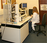
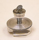
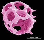
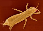
A scanning electron microscope or SEM has a scanning beam that sweeps across the specimen in lines and electrons emitted from the sample are collected by an objective, forming a 3D image of the sample. The specimens usually have to be coated with a thin layer of gold and are then mounted on to a special stub (illustrated below). Images are produced in black and white but can be false-coloured later. Please note the images presented here are low resolution images and are copyright to the originators; for a high resolution copy and permission to use them please apply to Dave Furness. |
|
|
|
|

|
Contact us
Central Electron Microscope Unit
School of Life Sciences
Keele University
Staffordshire
ST5 5BG
Professor Dave Furness
Director of the EM Unit
Email : d.n.furness@keele.ac.uk
Tel : 01782 733496
Simon Holborn
Email : s.c.holborn@keele.ac.uk
Tel : 01782 733484.


