Biomechanics and Biomedical Engineering
We carry out research in regenerative medicine and tissue engineering applications, aneurysm rupture in arteries, physiological flow in the lungs and fluid flow dynamics in mechanical thrombectomy devices. For more information on PhD opportunities, please see here.
Cell-based therapy (using autologous cartilage, bone and stem cells) is used mainly for the treatment, repair and regeneration of small areas of cartilage and bone damage resulting from accidental injury (e.g., to the knee joint) or tissue degeneration (e.g., in arthritis and osteo-arthritis). This therapy is being considered as a promising and viable alternative to artificial implants in large tissue regeneration such as the knee and hip. It could potentially improve the quality of life for the millions of arthritis and osteoarthritis sufferers worldwide. However, its benefit has not yet been clinically proven and is still only in the research trial phase. One of the main obstacles that clinicians face is that it is difficult and not feasible to continuously monitor the repair process following surgical insertion into the patient’s knee joint, for example. Hence, many details of the repair process are unknown. Our research focuses on developing mathematical models to enable better understanding of the repair process in humans.
We developed a mathematical model of cartilage regeneration after cell therapy. This model has enabled much better understanding of the repair process and has addressed some fundamental questions that are very useful in guiding practitioners of this cell-based therapy. Some of the highlights include showing the existence of an optimal initial seeding of cells which could benefit the repair process, the timeframe of regeneration and the fact that using stem cells alone is no better than using chondrocytes. These are in line with clinical studies performed at the Robert Jones and Agnest Hunt Othropaedic Hospital (RJAH), Oswestry, which have taken a timescale of over 5 years for this understanding to be gained. In this respect, our model provides a quick and cost effective way to realise the efficacy of this therapy.
- A mathematical model of cartilage regeneration after cell therapy, Lutianov, M., Naire, S., Kuiper, J.H. and Roberts, S. 2011, J. Theoretical Biology, vol. 289, pg. 136-150.
Subsequent research activity has focussed on including cell-to-cell interactions between the chondrocytes and cartilage cells into our model. We have shown that co-implantation of both chondrocytes and stem cells could be more beneficial to the repair process than implanting each individually
- A mathematical model of cartilage regeneration after chondrocyte and stem cell implantation - II: The effects of co-implantation. Campbell, K, Naire, S. and Kuiper, JH. 2019. Journal of Tissue Engineering, vol. 10. Clinical studies at RJAH currently use 50:50 ratios.
Another key area related to this therapy which is being intensely researched by biologists and clinicians is the role of certain growth factors in modulating cartilage regeneration. These growth factors are shown to result in particular cell-to-cell interactions which can be beneficial to the regeneration process at early time. We have included the role of growth factors into our model and have shown its benefit in enhancing cartilage at early time.
- A mathematical model of cartilage regeneration after chondrocyte and stem cell implantation -I: The effects of growth factors. Campbell, K, Naire, S, Kuiper, JH. Journal of Tissue Engineering, vol. 10.
Current research is focussed in understanding the mechanisms underlying osteochondral regeneration of a cartilage-bone defect. This understanding is crucial for the success of cell-based therapies for osteoarthritis sufferers and hence has huge potential impact to improve the quality of their lives. The underlying mechanisms of osteochondral regeneration are unknown and this research will be the first to develop this.
The above fundamental research directly benefits the actual practitioners of this cell-based therapy, i.e, the clinicians. Our mathematical models will enable better understanding of cell-based therapies in the regeneration of chondral and osteochondral which will hugely benefit the clinicians. This enhanced knowledge will guide the clinicians in making this therapy a success for improving the lives of millions of arthritis and osteoarthritis sufferers worldwide.
Collaborator
Dr Jan Herman Kuiper, ISTM, Keele University and Robert Jones and Agnes Hunt Orthopaedic Hospital in Oswestry.
The research was funded through the 3ME: Modelling Methods in Medical Engineering initiative (Michael Lutianov) and a Keele University PhD Studentship (Kelly Campbell).
Suction (aspiration) thrombectomy is a ground-breaking interventional procedure for removal of blood clots from the brain of patients suffering a stroke. While clinical trials have shown its effectiveness beyond any reasonable doubt, there are still gaps in understanding of the interaction between the suction, blood flow and clot deformation during the suction thrombectomy procedure. These are barriers to viability of this procedure for the large number of stroke patients who could benefit from this treatment.
A computational fluid dynamics model of a catheter suction device within an open and blocked blood vessel was used to simulate flow characteristics using the OpenFOAM CFD software package. The CFD model predicted that in an open vessel blood is drawn from upstream of the catheter tip (see panel B in figure) while in a blocked system blood is drawn from the vessel proximal to the device tip with no traction on the blockage distal of the tip (see panel A in figure). Corresponding in vitro benchtop experiments were performed at Keele University’s Institute of Science and Technology (ISTM) that confirmed the predictions of the CFD model. In the blocked vessel, suction had no effect on the blockage unless the tip of the catheter was in direct contact with it.
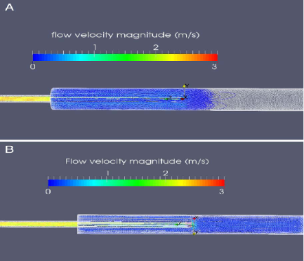
Results from the CFD modelling of the flow in a blocked vessel (top) and open vessel (bottom).
The experiments and CFD modelling suggested that suction is only effective if the catheter tip is in direct contact with the blockage. If the catheter tip is not in contact with the blockage, then blood is drawn from the vessels proximal of the blockage which could affect collateral blood flow and the successful outcome of the thrombectomy procedure in vivo.
Our findings contributed to a change in suction thrombectomy technique followed by clinicianal interventionalists. This demonstrates the potential of combining experimental and theoretical modelling to inform practice.
- In vitro experiments of cerebral blood flow during aspiration thrombectomy: potential effects on cerebral perfusion pressure and collateral flow, Lally F, Soorani M, Woo T, Nayak S, Jadun C, Yang Y, McCrudden J, Naire S, Grunwald I, Roffe C. 2016, J Neurointerv Surg, vol. 8(9), 969-972.
Current research is focussed in combining in vitro experiments with more advanced CFD modelling to consider the full 3-way interaction between the blood flow, clot deformation and suction as the clot is sucked out of the arterial network during the thrombectomy procedure.
Researchers involved
- John MCCrudden – MSc project in Biomedical Engineering, ISTM 2015.
- Riccardo Marconi – MSc project in Medical Technology, ISTM 2020.
Collaborators
- Dr. Christine Roffe (Consultant in Stroke Medicine at the Royal Stoke University Hospitals North Midlands (UHNM) NHS Trust and Honorary Professor at ISTM)
- Professor Ying Yang (Keele)
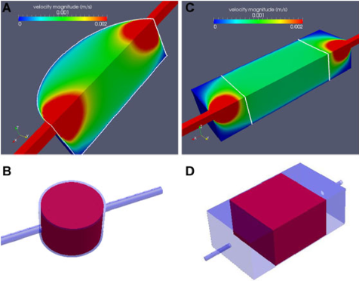
Perfusion bioreactors are commonly used in different tissue engineering applications because of their consistent distribution of nutrients and flow-induced shear stress within the tissue-engineering scaffold. A widely used configuration uses a scaffold with a circular cross-section enclosed within a cylindrical chamber and inlet and outlet pipes which are connected to the chamber on either side through which media is continuously circulated (see left panel B). However, fluid-flow experiments and Computational Fluid Dynamics (CFD) simulations have shown that the majority of the flow perfuses through the centre (see left panel A). This pattern creates stagnant zones in the peripheral regions as well as in those of high flow rate near the inlet and outlet (“hot-spots”). This non-uniformity of flow and shear stress, owing to a circular design, results in limited cell proliferation and differentiation in these areas.
In our study on Modeling and design of optimal flow perfusion bioreactors for tissue engineering applications, we challenged this design paradigm and provided an alternate design based on a rectangular bioreactor system (see panel D). CFD simulations within the rectangular bioreactor are shown to overcome the above flow limitations in the circular design (see panel C).
- Modeling and design of optimal flow perfusion bioreactors for tissue engineering applications, Hidalgo-Bastida LA, Thirunavukkarasu S, Griffiths S, Cartmell SH, Naire S. 2012. BIOTECHNOLOGY AND BIOENGINEERING, vol. 109(4), 1095-1099
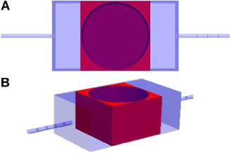
However, from a clinical point of view, a rectangular scaffold is not desirable since surgical techniques do not include drilling rectangular holes to implant tissue engineered construct at damaged tissue sites. Practically this issue can be resolved in two ways. Either a circular cross-section scaffold is used but enclosed within a rectangular porous holder (see right panel A). This holder should ideally be made of a similar material as the scaffold and pre-treated to deter cell growth from the scaffold to the holder. The second alternative is to use a square scaffold and then cut-out a cylindrical section once the flow perfusion is completed (see right panel B). However, the risk of damaging the cells during this needs to be addressed by adequate processing of the construct. In any case, both alternatives can satisfy the clinical requirement of a circular cross-section.
This work was part of Sundar Thirunavukkarasu’s MSc project in Biomedical Engineering and funded through the 3ME: Modelling Methods in Medical Engineering initiative.
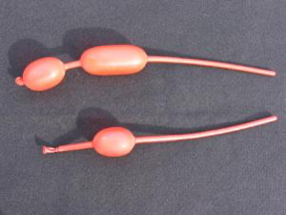
An aneurysm is a localized, blood-filled balloon-like bulge of a blood vessel. As the size of an aneurysm increases, there is an increasing risk of rupture, resulting in severe hemorrhage, other complications or even death.
The geometrical similarity between an arterial aneurysm and a localized bulge in tubular party balloons is obvious (see picture below). We are hoping to use our knowledge of the bulging process associated with party balloons to shed light on the initiation process of aneurysms.
Our first observation is that although party balloons always suffer localized bulging when inflated, healthy arteries do not. What is then the design principle for bulge-resistant arteries, and under what pathological changes can aneurysms occur? Some of the questions that we have answered so far are:
(1) Why do party balloons bulge when inflated and at what pressure do they bulge?
[1] Post-bifurcation analysis of a thin-walled hyperelastic tube under inflation, IJNM (2008)
[2] An experimental study of localized bulging in inflated cylindrical tubes guided by newly emerged analytical results, JMPS (2019)
[3] Weakly nonlinear analysis of localized bulging of an inflated hyperelastic tube of arbitrary wall thickness, JMPS (2020)
(2) Can we improve bulge resistance by increasing wall thickness?
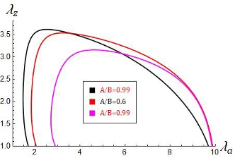
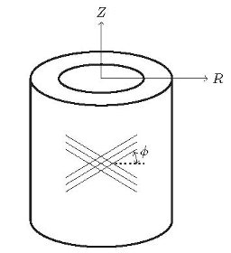
(3) Arteries are multi-layered and fibre-reinforced. How do fibre reinforcement and optimized fibre orientation help prevent bulge formation?
Collaborators
- Professor Shibin Wang, Linan Li, Tianjin University, China
- Dr Juan Wang: University of Shanghai for Science and Technology, China

People
Projects
- Physiological fluid mechanics, regenerative medicine / tissue engineering (Dr. S. Naire)
- Continuum-mechanical modelling of the formation and rupture of aneurysms in human arteries (Professor Y. Fu)
PhD Students
Current
- Dominic Emery (Supervisor: Yibin Fu)
Recent
- Hani Alahmadi (Supervisor: Shailesh Naire )
- Kelly Campbell (Supervisor: Nair Shailesh)
- Ali Althobaiti (Supervisor: Yibin Fu)
- Geethamala Francis (Supervisor: Yibin Fu)