Regenerative Medicine - Equipment
OS Equipment
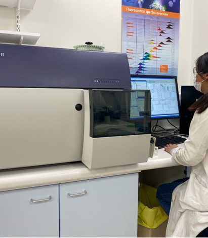 The BD FACSCanto II system has the versatility to meet demanding research requirements. Sample introduction can be accomplished in a single tube or via the BD™ fully automated carousel. BD FACSDiva™ software controls the setup, acquisition, and analysis of flow cytometry data. The flow cytometer has three solid state lasers, a red, blue and violet laser, which means up to 8 flurophores can be analysed simultaneously on a single cell.
Funded by Versus Arthritis
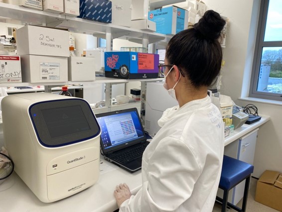 The QuantStudio 3 from applied biosystems is a 96-well 4-colour qPCR machine capable of a wide range of assays. Its main benefits are high sensitivity and wide range, detecting changes in target quantity as small as 1.5-fold in single-plex reactions and obtaining 10 logs of linear dynamic range. Another benefit of this system is the low sample and reagent requirement. QuantStudio Design and Analysis desktop software can design experiments and analyse results from the computer or from the machine itself.
Funded by HEFCE
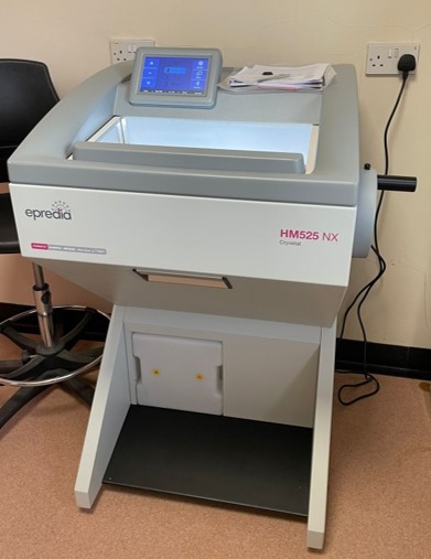
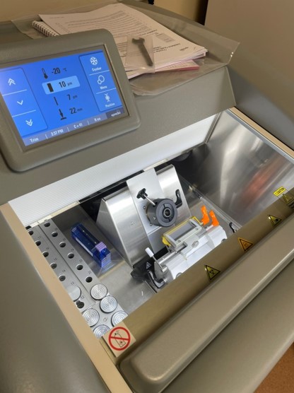
Our brand new Epredia™ HM525 NX cryostat is a high-performance routine cryostat with intuitive software and touchscreen for simple, efficient operation with a user-friendly and ergonomic design. The cryochamber cools to -35°C and features 27 cooled specimen positions, including four fast-freeze stations (cooled to -60°C) to keep up with heavy workloads. A reliable stepping motor technology delivers reproducible section thickness, ranging from 1µm to 500µm. It includes a UV disinfection mode to protect against occupational exposure to biological pathogens and surface contamination.
Funded by the Orthopaedic Institute
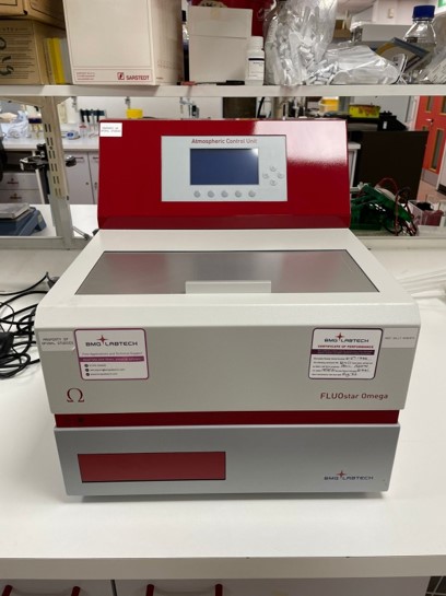 The FLUOstar® Omega is a multi-mode microplate reader with six detection modes: fluorescence intensity (including FRET), luminescence (including BRET), UV/vis absorbance, time-resolved fluorescence, TR-FRET, and AlphaScreen®/AlphaLISA®.. It utilizes an ultra-fast UV/vis spectrometer or filters for absorbance as well as highly sensitive filters for all other detection modes. The LVis Plate is suitable for quick and easy low-volume concentration measurements of up to 16 DNA, RNA, protein samples, or spectral scanning. The Atmospheric Control Unit (ACU) enables live cell-based applications, from standard proliferation to hypoxic or cytotoxicity assays by independently regulating both O2 and CO2 gas levels within the microplate reader chamber.
Funded by HEFCE
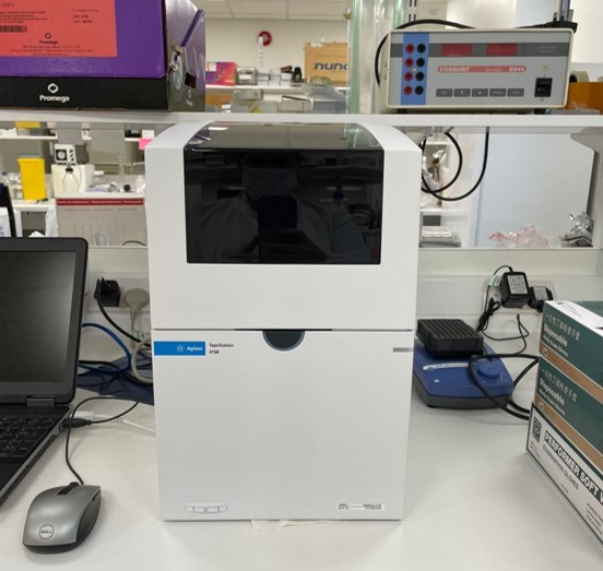 The Agilent 4150 TapeStation System is a unique all-in-one platform that includes instrumentation, data processing software, and reagents for the analysis of quantity and integrity of DNA and RNA by proprietary small-scale gel electrophoresis. The system is fully automated, using only 1-2ul sample per run and can analyse up to 16 samples at once with results available in under 20 minutes. The Tapestation provides reliable and reproducible quality control data such as Integrity standards for RNA (RNA integrity number equivalent, RINe) and genomic DNA (DNA Integrity Number, DIN) required for downstream analyses such as next-generation sequencing.
Funded by the Orthopaedic Institute
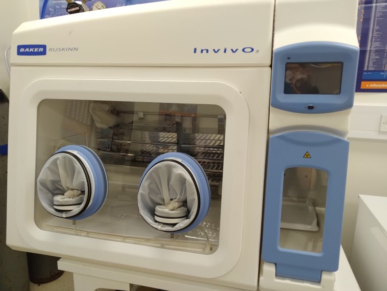
The InvivO2 Cell Culture Workstation allows you to establish a cell culture environment that reproduces natural physiological conditions. Oxygen, carbon dioxide, temperature and humidity can be customised to your culture and experimental needs. Such parameters are monitored constantly and direct-hand access allows you to conduct cell culture without leaving these optimal conditions to ensure precise results.
Funded by the Orthopaedic Institute
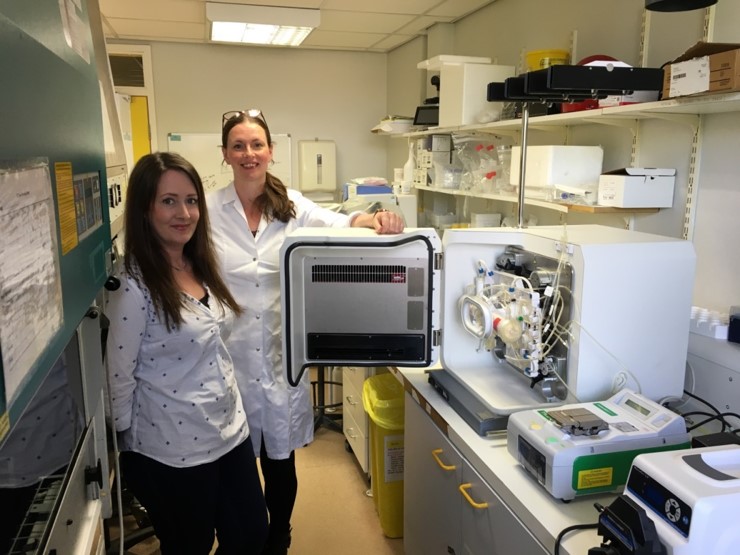
The Quantum Cell Expansion System is an automated platform designed to simplify the open, labour-intensive tasks associated with cell culture. This hollow fibre bioreactor can be used for adherent and non-adherent large-scale cell culture. Cells can be maintained under constant perfusion in normoxic and hypoxic conditions. The bioreactor consists of 11.5k hollow fibres with semi-permeable membranes. This equates to a surface area of 2.1m2. The system is best suited to the loading of high cell numbers (i.e. millions), which can more effectively utilise all the fibres.
Funded by the Medical Research Council
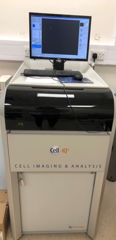 Cell-IQ® is a fully integrated, automated cell culture and analysis system for kinetic cell studies. The system comprises of an incubator and imaging hardware platform with powerful intuitive software for data capture and processing. Following a simple set up, the Cell-IQ software enables cells to be monitored continuously via the Imagen™ program, whilst the Analyser™ software automates the process of quantitation measuring multiple cellular parameters from phase contrast and fluorescence images. The unique ability to quantify cells/cell phases directly from the phase images based their morphology alone makes Cell-IQ a truly label free technology.
Funded by the Orthopaedic Institute
GHRC equipment
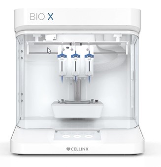
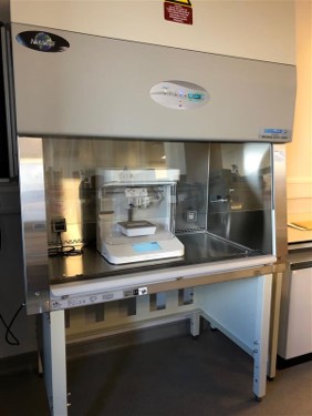 The Bio X is a user friendly and flexible 3D biobrinter which bioprints with cells using bioink to provide a temporary or permanent support to the cells while they produce their own extracellular matrix. Numerous bioinks are available depending on cell type, and multiple different printing styles are possible using the different print heads, these include pneumatic, thermoplastic, temperature controlled, electromagnetic droplet and a syringe pump print head. These can be used simultaneously in 3 print head holders. 365nm and 405nm exchangeable photocuring modules are also available for crosslinking.
Funded by the EPSRC
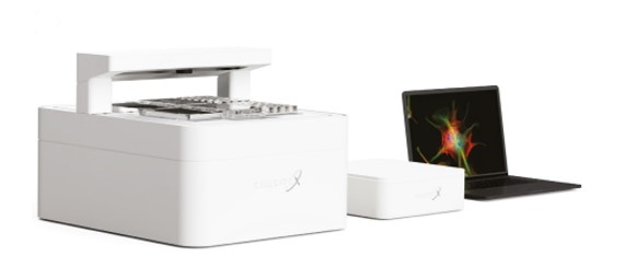 The CellCyte is a high-throughput live cell imaging system allowing cells to be imaged in real time and at time points defined by the user. 6 plates can be run simultaneously using 6 - 96 well plates in each slot, Multiple images can be taken per well. A x10 objective with auto focusing is used to capture brightfield images as well as fluorescence images using 3 fluorescence channels, blue, green and red. There is also an analysis package which can be used to determine confluency, cell count and nuclei count.
Funded by the EPSRC
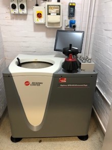 The Optima XPN ultracentrifuge allows samples to be spun at up to 70,000rpm with 10 acceleration profiles and 11 deceleration profiles. Up to 1500ml of sample can be spun at once at a temperature range between 0-40°C. A fixed angle rotor as well as a swinging bucket rotor are available. The inbuilt software can be used to quickly perform common calculations and conversions in order to optimize protocols and save time and samples.
Funded by the UKRI
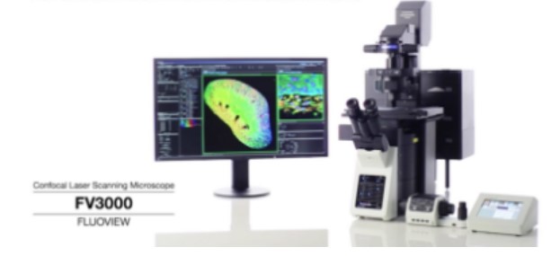 The FV1200 confocal microscope enables a wide range of imaging modalities, including macro-to-micro imaging, super resolution microscopy, and quantitative data analysis. 4 lasers are available (405nm, 473nm, 561nm, 635nm) which are individually adjustable to optimize signal detection for each fluorophore. Numerous objectives are available including a 30X silicone lens which has a refractive index close to living tissue enabling high resolution observation deep inside tissue with minimal spherical aberration.
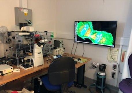 The Nikon Eclipse Ti microscope with environmental chamber and automatic stage allows cells to be analysed in real time, and time lapse studies to be carried out in optimal conditions. The microscope features a 4.2 megapixel, cooled, monochrome camera, plus a pE-300 LED light source for fluorescence imaging with DAPI, FITC, TRITC, and far-red filters. The 20x and 40x fluorescence objectives are compatible with thick plastic bottom flasks and well plates and enable high-resolution phase-contrast and fluorescence observations. They also feature a correction collar for spherical aberration correction.
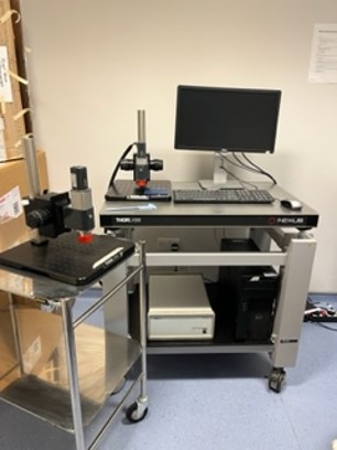 The Telesto-II OCT is capable of producing high resolution, cross sectional images of materials including biological tissue, bridging the gap between confocal and ultrasound imaging. It utilises a centre wavelength of 1300nm providing around 3.5mm imaging penetration with 5.5µm axial resolution. Both 2D and 3D images can be taken to display cross sectional images in real time, constructed into a 3D volumetric image for 3D scans.
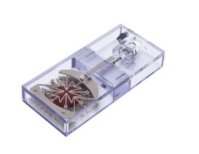 The MCB1 bioreactor generates radial motion patterns on a 24-pin mounting ring. 35mm diameter membranes, scaffolds, and tissues are then mounted to this ring and biaxially stretched up to 20% according to the specified protocol. The entire assembly can be sterilized and is suitable for long-term cell culture in a laboratory incubator. A wide variety of materials including silicone membranes, decellularized tissues, hydrogels, electrospun materials, and more can be used in the system.